The Muscular System 101: Understanding Muscle Anatomy & Regeneration
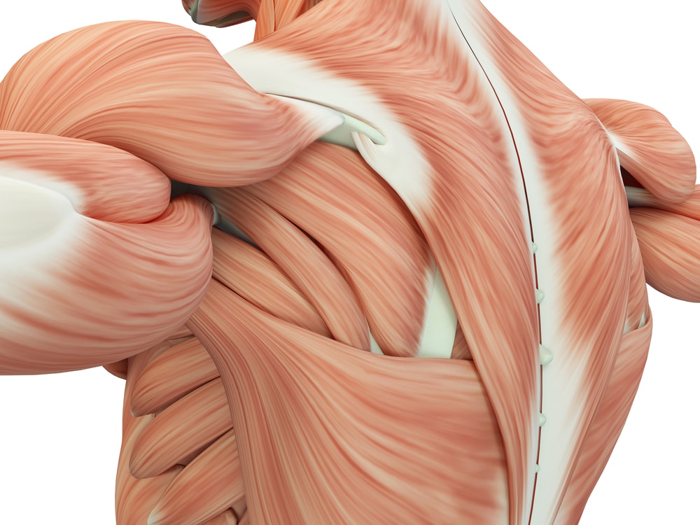
There are three kinds of muscle in the human body — but the most relevant for the purpose of this discussion is the skeletal muscle. The other two are smooth muscle (found in the internal organs and lining of the arteries), and cardiac muscle (a specific type of muscle only found in the heart).
The cells that make up skeletal muscle are uniquely suited to their job. They have more mitochondria (the powerhouse of the cell) than most other cells in the body. This is meant to help them meet the high energy demands of the muscles.
Muscle cells are also long and cylindrical, helping them form long fibers.
Each muscle cell contains functional units called sarcomeres — which are the part of the muscle that contracts and expands to allow for movement. Proteins called actin and myosin contained in the cells are responsible for expanding and contracting the muscle tissue to achieve movement using calcium and adenosine triphosphate (ATP).
When a nerve stimulates muscle cells with acetylcholine, (i.e. when you tell your arm to move) calcium is pumped into the cell, which allows actin and myosin to interact, causing the cell to contract (shorten). This step requires a lot of energy — in the form of ATP, produced in the mitochondria of the cell.
When the nerve stops activating the muscle cells, calcium is pumped back out of the muscle cell and phosphate (a form of transferable energy) dislodged, causing the muscle to expand again (lengthen).